The Potential to Regenerate the Retina Using Stem Cells
Photoreceptors are light-sensitive cells that line the back of the eye like the pixels of a digital camera and allow us to see. In the USA and Europe, most people who are registered blind have suffered degeneration or loss of these cells. Writing in the journal Nature, however, researchers at the UCL Institute of Ophthalmology in London describe a method of successfully transplanting photoreceptor cells ? the key is getting the timing right.
MacLaren R.E., Pearson R.A., MacNeil A., Douglas R.H., Salt T.E., Akimoto M., Swroop A.S., Sowden J.C., Ali R.R. (2006) Retinal repair by transplantation of photoreceptor precursors. Nature 444:203-07.
Loss of photoreceptor cells occurs in age-related macular degeneration or genetic eye diseases, such as retinitis pigmentosa. Although some new treatments may be effective in the early stages of these diseases, loss of photoreceptor cells has always been considered to be irreversible. Like other parts of the adult brain, fully mature photoreceptors cannot be transplanted and recent research has focussed on the potential to regenerate the retina using stem cells. It is very difficult however to get stem cells to turn into photoreceptors. The researchers in London therefore tried a different approach and transplanted semi-mature cells that were about to become photoreceptors, known as photoreceptor ‘precursor’ cells. The researchers found that the precursor cells, having gone much further along the line of photoreceptor development than stem cells, were able to complete their maturation after transplantation to become photoreceptors. Some of the transplanted photoreceptor cells were able to make connections and restore vision. To assess vision, the cells were transplanted into the retina of mice which did not have any functioning rod photoreceptors. Rods are the light sensitive cells used for night-time vision (cones are used during the day) and loss of rod cells also occurs in the disease known as ‘retinitis pigmentosa’, which affects patients. The researchers assessed vision in the mice by looking at how the iris constricted when light was shone in the eye. They found that the pupils of the transplanted eyes could respond to low levels of light after transplantation and that the degree of pupil constriction was directly correlated to the number of successfully transplanted rod photoreceptor cells. In this way, the mice that previously could not see at this level had their pupil reflex restored. In a mouse, this is about as close as one can get to saying that the mice can see.
In order to translate this in to a clinical treatment (which according to the researchers is some years off) more cells will be required for transplantation. Probably millions would be required and in humans these would need to be the cone type of photoreceptor cells rather than rods, because our sight is optimised for daytime use. Possibly adult stem cells rather than embryonic stem cells might be the way forward.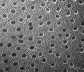 Fig 1 Photoreceptors in the human retina
Microscopic cross section of photoreceptors in the human retina. Photoreceptors generate electrical signals in response to light. The small circular cells seen here are rod photoreceptors, which are used for night vision and the larger cells are cones, which are used for daytime and colour vision. Each human eye contains about 125 million rods and 6 million cones. (Picture courtesy of Dr Peter Munro, UCL Institute of Ophthalmology, London EC1V 9EL)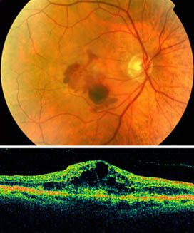 Fig 2 Macular degeneration
A photograph of the back of the eye in a patient with age-related macular degeneration. The image above shows blood and degeneration in the central part of the retina. The white circle to the right is the optic nerve, corresponding to the blind spot. Elsewhere the retina is lined with millions of light sensitive photoreceptor cells. The image below is an optical section through the central part of the retina above. The normally smooth contour of the retina has large black areas where photoreceptors have been lost due to the macular degeneration.
(Picture courtesy of Dr Robert MacLaren, Moorfields Eye Hospital, London EC1V 2PD) Fig 3 Transplanted rod and cone
A microscope image showing transplanted photoreceptors from the paper by MacLaren et al. published this week in Nature. The rod photoreceptor on the left can be distinguished in shape from the cone photoreceptor on the right. Only the transplanted cells are fluorescent green due to a genetic label, which clearly distinguishes them from the host photoreceptors which are coloured blue.
(Picture from paper by MacLaren et al., courtesy of Nature Publishing Group)
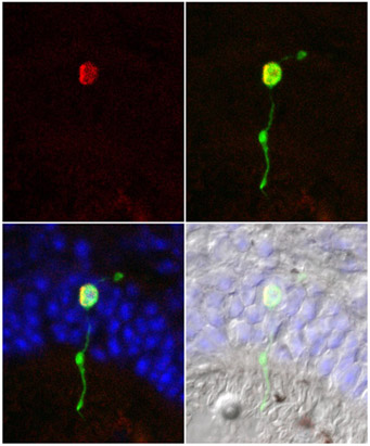 Fig 4 Montage showing nucleus
A montage showing a single transplanted photoreceptor cell in the host retina. The red circle represents the cell nucleus which was labelled during cell division prior to transplantation. The transplanted cell is also identified by green fluorescence, against the host retina cells which are blue. By identifying cell division using this red label, the authors concluded that cells must undergo a final division before becoming transplantable.
(Picture from paper by MacLaren et al., courtesy of Nature Publishing Group)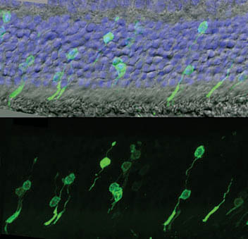 Fig 5 Transplanted photoreceptors
Transplanted photoreceptor cells can be seen by green label in these images. The picture above shows the whole retina with the host retinal cells labelled blue. In the picture below, only the green channel is shown to highlight the shape of the transplanted cells. The round nucleus and long tails are the typical shapes of rod photoreceptor cells.
(Picture from paper by MacLaren et al., courtesy of Nature Publishing Group)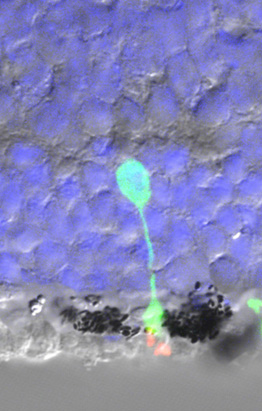 Fig 6 Transplanted photoreceptor and pigment
A high power image showing the position of a single transplanted photoreceptor cell in contact with dark pigment granules of the host retina. These granules absorb stray light and help to improve the image quality. The light sensitive molecules can also be seen in red in this image. The fluorescent green colour is a genetic marker that confirms the identity of the transplanted cells. The host retinal cells are shown in blue.
(Picture courtesy of Dr Robert MacLaren, Moorfields Eye Hospital, London EC1V 2PD)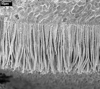 Fig 7 Cross section of the human retina
This image shows the densely packed photoreceptors in the human retina. The long tails contain molecules that generate electrical impulses in response to light. Most of the cells here are rod photoreceptors, which are used for seeing in the dark, but two chubby cells can be seen in the centre of the image. These are cone photoreceptors, used for colour vision.
(Picture courtesy of Dr Peter Munro, UCL Institute of Ophthalmology, London EC1V 9EL)


I'm a biology teacher in France and I need information about the 1st picture :
From witch part of the retina comes this picture ?
Thanks for your answer.
Laurence Durand
[Response] Bonjour Laurence, It is the macular region of the human retina just outside the fovea. The large cells are cones and you can see that there are more rods in this region. In the fovea they would all be cones. I hope this helps. Robert
[Response] There is unfortunately no effective treatment for any retinal disease using stem cells at present. Of course, this may change in the future. I have several patients with ROP and they live perfectly normal lives and because the sight loss occurs very early, they learn to adapt to it. It is important to accept the diagnosis and give your child all the love and support necessary, without focussing on something that is wrong that might be cured.
Any advise will be deeply appreciated.